
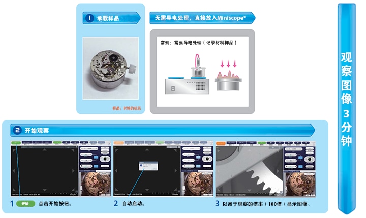
◆ Observe the image for 3 minutes. The required data can be obtained quickly and reports can be made.
Observation and analysis flexibility
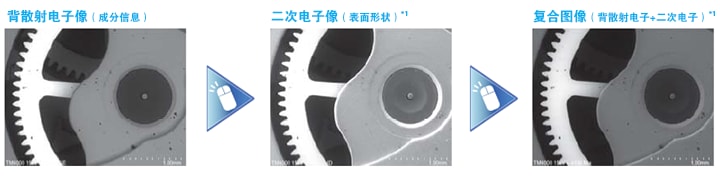
Obtain all kinds of data automatically. Switch quickly!
Quickly obtain the element distribution map*2
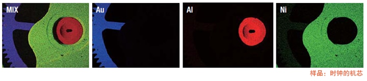
*1 The function of TM4000PlusⅡ. *2Optional
With Camera Navi*, it’s that simple
Camera Navi image allows you to easily find the field of view, the distribution map (MAP) function supports your observation throughout the process
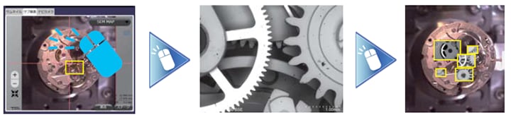
*Optional: Camera Navi system
You can also watch the video
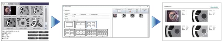
Simple and fast operation
It only takes 3 minutes to observe the image.
You can quickly observe the image and export the test report.
Report Creator allows you to easily create reports
You can create reports in Microsoft Word, Excel, and PowerPoint formats by simply selecting images and templates
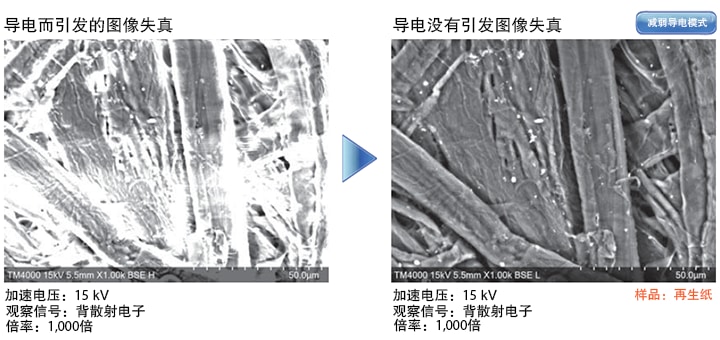
◆ Even the insulation samples can be observed directly without pretreatment.
"Static Reduction Mode" can suppress static electricity
For samples that are prone to static electricity, you can use the "static electricity reduction mode" to observe in a state where static electricity is suppressed.
Simply click on the software with the mouse to switch to the "static reduction mode".
Various observations can be performed under low vacuum conditions
For samples that are prone to static electricity, such as powder or water, the observation can be combined with its purpose.
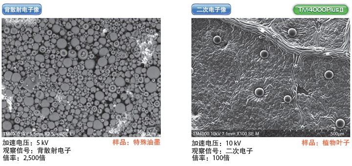
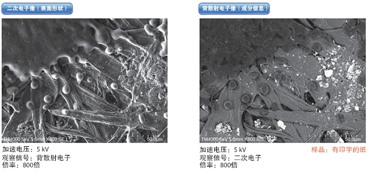
◆ The secondary electron image (surface shape) can be observed under low vacuum conditions.
No pretreatment is required, and the surface of insulators and water-containing and oily samples can also be observed
Not only to observe traditional conductive samples, but also to observe insulators and water-containing and oily samples without pretreatment. It can quickly switch between the secondary electron image and the backscattered electron image.
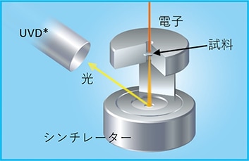
High sensitivity low vacuum secondary electron detector
Using high sensitivity low vacuum secondary electron detector (UVD). By detecting the light generated by the collision between electron beams and residual gas molecules, images with secondary electron information can be observed. In addition, by controlling the detector to detect the light generated by the electron irradiation, CL information (UVD-CL: Image with CL information) can be obtained.
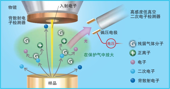
The detection principle of high sensitivity low vacuum secondary electron detector
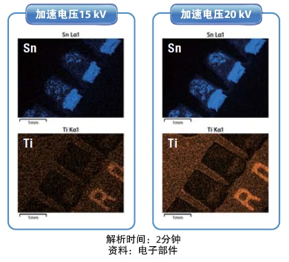
◆ Support acceleration voltage 20 kV
TM4000Ⅱ/TM4000Plus II can support an acceleration voltage of 20 kV.
With EDS analysis (optional), higher counting rate analysis can be performed.
By accelerating voltage of 20 kV, high S/N of the EDS element distribution map is realized
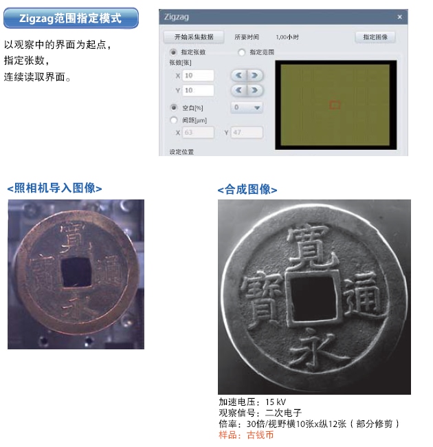
◆ Multi Zigzag (optional)
It can realize SEM observation in a wide area.
With automatic motor stage, low magnification, high precision, and large-scale observation and analysis can be realized.
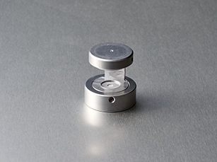
◆ EM sample table (optional)
Easily observe STEM images
With the newly developed STEM sample stage and high-sensitivity low-vacuum secondary electron detector (UVD), it can easily observe small-magnification STEM images.
Easily observe film samples and biological samples.
* UVD is an accessory on TM4000Plus II.
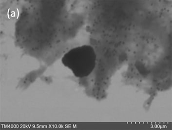
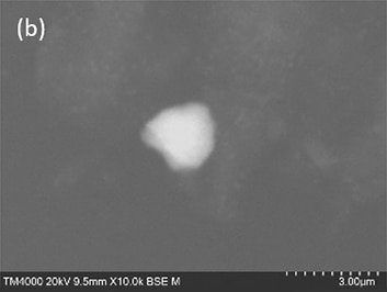
Sample: Abrasive
Accelerating voltage: 20 kV
Observation signal: (a) STEM image, (b) backscattered electron image
Magnification: 10000 times
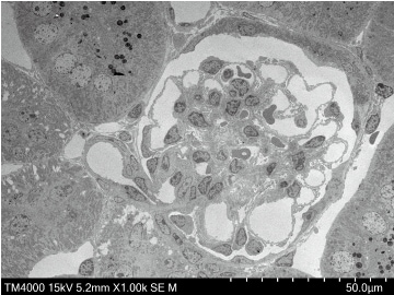
Sample: Mouse Kidney
Accelerating voltage: 15 kV
Observation signal: STEM image
Magnification: 1000 times
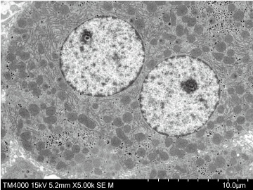
Sample: Mouse liver
Accelerating voltage: 15 kV
Observation signal: STEM image
Magnification: 5000 times