
◆ Core Concept
1. Use excellent imaging technology
SU7000 can quickly acquire a variety of detection signals from the wide-view full-view image to the fine surface structure. The newly designed electronic optical system and detection system enable the device to receive signals such as secondary electrons and backscattered electrons at the same time, and users can obtain more comprehensive sample information in a short time.
2. Adopt multi-channel imaging technology
As users' demand for imaging continues to grow, SU7000 has added several new detectors and introduced corresponding imaging functions. SU7000 can display and save up to 6 channels of signals at the same time. Achieved to obtain sample information.
3. Support different shapes of samples and multiple observation methods
・Large sample observation
・Low vacuum observation
・Ultra low temperature observation
・Real-time observation
The required sample chamber and vacuum system are very complete, and the observation methods are also readily available.
4. Support micro-nano analysis
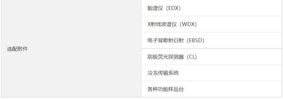
Using a Schottky electron gun, the beam current can reach 200 nA, which is suitable for various micro-nano analysis. The sample chamber shape and interface settings support EDX analysis, EBSD, cathodoluminescence analysis, etc. The interface equipment can meet various special needs through optional accessories.
◆ Imaging capability
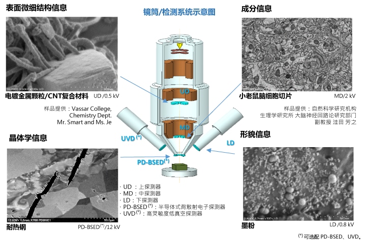
The detection system pursuing the large-scale information
As users' requirements for sample data become more diversified, higher requirements are placed on the detection system to capture more information in a short period of time. The SU7000 detection system can obtain information such as morphology, composition, crystallography, and luminescence more efficiently without changing the conditions such as WD. Realize faster and more comprehensive information collection.
Observe in the same condition
Sample: Metal rod wrapped with organic matter
Sample provided: Vassar College, Chemistry Dept.
Mr. Smart and Ms. Pineda
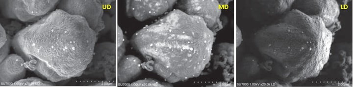
Application example of collecting signals simultaneously through UD, MD, and LD detectors (accelerating voltage: 1 kV)
The UD, MD, and LD detectors can collect signals at the same time, UD can obtain surface fine shape information, MD can obtain surface organic matter coating state information, and LD can obtain overall uneven topography information. Under the same conditions, information was obtained.
◆ Adopting GUI design with multi-channel display
Optimized signal display function
The imaging unit has also been upgraded in signal display.
image.png Users can choose different viewing angles and channel numbers according to observation requirements.
image.png A single screen can display up to 4 channels of signals at the same time.
image.png The navigation function (SEM MAP) can easily capture the CCD camera image showing the information in the sample compartment and the sample image taken by the camera, and quickly display the target image.
image.png When dual-screen display is used, a 1,280×960 pixel image can be displayed in two channels on one display screen, and the other display screen is dedicated to display signals, which can display up to 6 channels of signals at the same time.
High degree of freedom interface layout
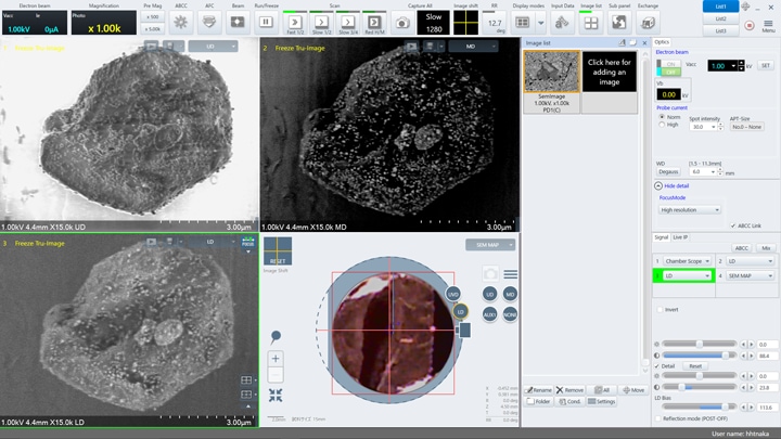
When using a single screen, you can display sample information in a single channel, dual channels, or four channels, as well as CCD camera images, sample stage images/SEM MAP images, etc. And the toolbar on the operation screen can be moved, added and deleted at will.
Support dual screen display
The 1st display is dedicated to display images, and the 2nd display is used to display the operation panel screen. The left picture above (1st display) is a picture of non-metallic inclusions in steel observed through SU7000. The picture clearly shows the images captured by the five detectors UD, LD, UVD, MD, and PD-BSED, as well as the SEM MAP image. The picture on the right (2nd display screen) shows the operation panel menu and the image history window on the same screen, and the interface layout is clear. While realizing real-time monitoring images, the operating performance has also been further improved.
◆ Extensible observation and analysis methods
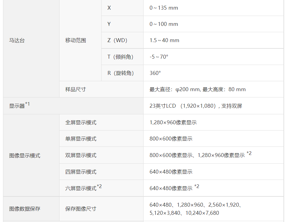
Sample compartment and motor stage
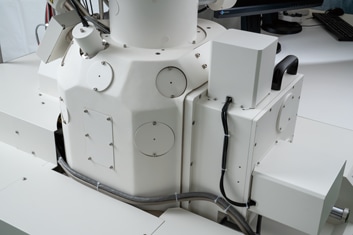
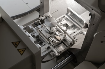
Appearance of sample bin Motor table appearance
Standard 18 accessory interfaces The movable distance of XY axis is 135×100 mm
The sample compartment can carry samples with a diameter of φ 200 mm and a height of 80 mm. Large opening design, standard 18 accessory interfaces. The sample stage can carry samples with a movement distance of 135×100 mm on the XY axis and a maximum weight of 2 kg. It is not a problem to carry large samples and functional sample stage.
Camera navigation function (*)
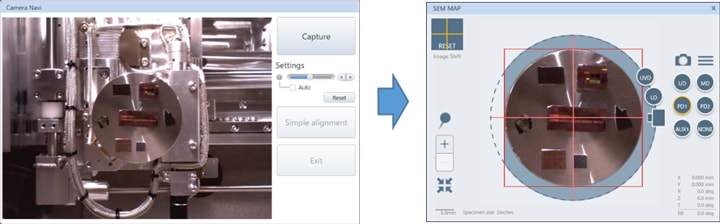
Take samples by camera Display the image captured by the camera on the SEM MAP screen for navigation
SU7000 can optionally be equipped with a navigation camera (*) to quickly lock the position to be observed. The camera is installed in the sample compartment. After the sample is inserted, the overall image of the sample can be taken semi-automatically. The image will be displayed in the navigation screen (SEM MAP) window, so the user can observe the image in the window to determine the position. The applicable size is φ 100 mm.
(*) Optional camera.
Detection system supporting dynamic observation
SU7000 can be used for dynamic observation of environmental changes. For low-vacuum observation, various detectors (**) such as PD-BSED (backscattered electron detector), UVD, MD, etc. can be used.
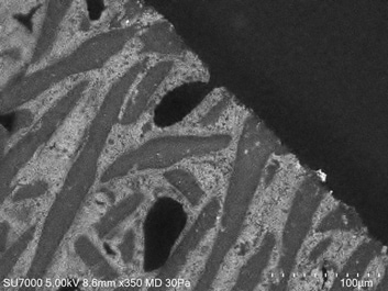
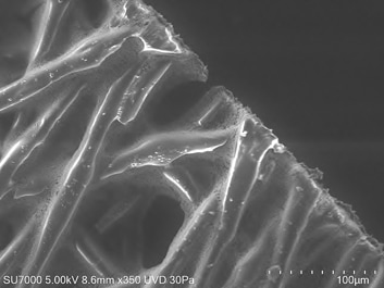
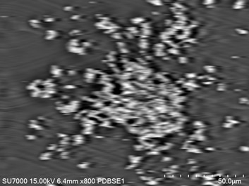
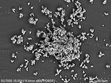
Detector selection for low vacuum environment
The picture on the left is through MD (backscattered electron), and the picture on the right is the metal oxide fiber observed by UVD (secondary electron) detector. The two pictures clearly show the dispersion state of oxides and the accumulation of fibers.
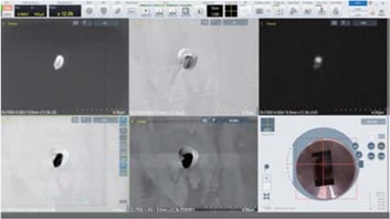
PD-BSED: Increased response speed
The image on the left is an image obtained by scanning once every two seconds with a normal response speed; the image on the right suppresses the drift of the image, which greatly improves the overall image quality. As the response speed increases, it is more suitable for In-Situ observation.
(**) PD-BSED and UVD are optional.

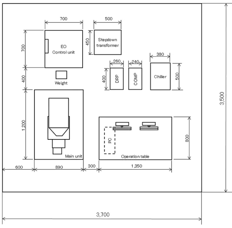
安装布局: