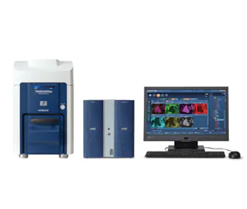
Scanning electron microscope is used to observe biological slices, biological cells, bacteria and living tissue culture, fluid precipitation, etc. Observation and research, at the same time can observe other transparent or translucent objects, powder, fine particles and other objects.
Biological microscopes are used for the observation of microorganisms, cells, bacteria, tissue culture, suspensions, sediments, etc. in medical and health institutions, colleges and universities, and research institutes. It can continuously observe the reproduction and division process of cells and bacteria in the culture solution. It is widely used in the fields of cytology, parasitology, oncology, immunology, genetic engineering, industrial microbiology, and botany.
Today, the editor will introduce the methods and specific steps of using biological microscopes:
1. Take the lens and place it
1. Hold the mirror arm with your right hand and the mirror holder with your left hand.
2. Place the microscope on the experiment table, slightly to the left (the microscope is placed about 7 cm from the edge of the experiment table). Install the eyepiece and objective lens.
Second, the light
3. Rotate the converter to align the low-power objective lens with the light hole (the front end of the objective lens and the stage should be kept at a distance of 2 cm).
4. Align a larger aperture at the light hole. Look into the eyepiece with your left eye (open your right eye for easy drawing at the same time later). Rotate the reflector to reflect the light into the lens barrel through the light hole. Through the eyepiece, you can see the bright white field of vision.
Three, observation
5. Put the glass slide specimen to be observed (it can also be made of thin paper sheet printed with "6") on the stage, and press it with the sheet clamp, the specimen should be facing the center of the light hole.
6. Rotate the coarse collimation screw to slowly lower the lens barrel until the objective lens is close to the slide specimen (look at the objective lens to prevent the objective lens from touching the slide specimen).

7. Look into the eyepiece with your left eye, and at the same time turn the coarse collimation screw in the opposite direction to make the lens barrel rise slowly until the object image is clearly seen. Turn the fine focus screw slightly to make the image of the object more clear.
8. Use of high magnification objective lens: Before using high magnification objective lens, you must first find the observed object image with low magnification objective lens and adjust it to the center of the field of view, and then turn the converter to change the high magnification lens. After switching to a high-power lens, the brightness in the field of view becomes darker, so generally choose a larger aperture and use the concave surface of the mirror, and then adjust the fine collimation spiral. The number of objects to be viewed decreases, but the volume becomes larger.
Fourth, organize
9. Scanning electron microscope, after the experiment, wipe the surface of the microscope clean. Rotate the converter, shift the two objective lenses to the sides, and slowly lower the lens barrel to a low position, and place the reflector vertically. Finally put the microscope into the mirror box and return it to its original place.