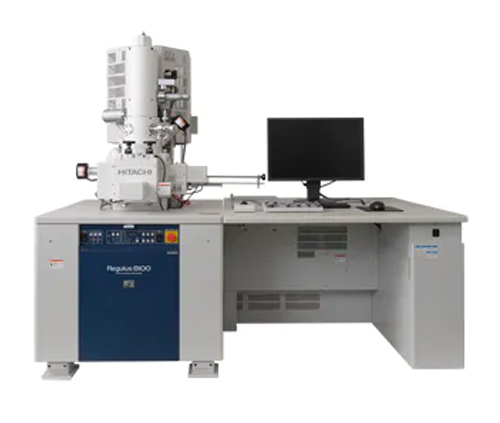
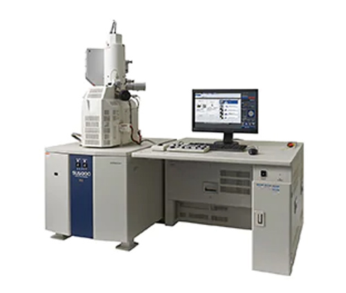
(1) Structure
1. Lens barrel
The lens barrel includes an electron gun, a condenser lens, an objective lens and a scanning system. Its function is to generate a very thin electron beam (about a few nm in diameter) and scan the electron beam on the surface of the sample while exciting various signals.
2. Electronic signal collection and processing system
In the scanning electron microscope sample chamber, the scanning electron beam interacts with the sample to generate a variety of signals, including secondary electrons, backscattered electrons, X-rays, absorption electrons, Auger electrons, and so on. Among the above signals, the most important is the secondary electrons, which are the outer electrons in the sample atoms excited by the incident electrons. They are generated in the region from a few nm to tens of nm below the sample surface. The generation rate is mainly determined by The morphology and composition of the sample. The commonly referred to as scanning electron image refers to the secondary electron image, which is the most useful electronic signal for studying the surface morphology of a scanning electron microscope sample. The detector for detecting secondary electrons (the probe of Fig. 15(2) is a scintillator. When electrons hit the scintillator, 1 generates light in it. This light is transmitted to the photomultiplier tube by the light pipe, and the light signal That is, it is converted into a current signal, and then through pre-amplification and video amplification, the current signal is converted into a voltage signal, and finally sent to the grid of the picture tube.

3. Electronic signal display and recording system
The image of the scanning electron microscope is displayed on a cathode ray tube (picture tube), and is photographed and recorded by a camera. There are two picture tubes, one is used for observation and has a lower resolution and is a long afterglow tube; the other is used for photographic recording and has a higher resolution and is a short afterglow tube.
4. Vacuum system and power system
The vacuum system of the scanning electron microscope is composed of a mechanical pump and an oil diffusion pump. Its function is to achieve a vacuum degree of 10 (4~10 (5 Torr) in the lens barrel. The power supply system supplies the specific power required by each component.

(2) Working principle
The electron beam from the cathode of the electron gun with a diameter of 20(m~30(m) is affected by the accelerating voltage between the anode and the cathode. Needle. Under the action of the scanning coil on the upper part of the objective lens, the electronic probe scans the surface of the sample in a raster shape and excites a variety of electronic signals. These electronic signals are detected by the corresponding detector, amplified and converted, and become voltage signals. Finally, it is sent to the grid of the picture tube and modulates the brightness of the picture tube. The electron beam in the picture tube is also scanned in a raster shape on the phosphor screen, and this scanning movement is strictly synchronized with the scanning movement of the electron beam on the sample surface, so that the contrast is obtained. The scanning electronic image corresponding to the intensity of the received signal. This image reflects the topographical characteristics of the sample surface. Section 2 Scanning Electron Microscopy Biological Sample Preparation Technology Most biological samples contain water and are relatively soft. Before scanning electron microscope observation, the samples should be processed accordingly. The main requirements for SEM sample preparation are: as far as possible, the surface structure of the sample should be preserved without deformation and pollution, and the sample should be dry and have good electrical conductivity.