
The imaging principle of electron microscope and optical microscope is basically the same, the difference is that the former uses electron beam as light source and electromagnetic field as lens. In addition, because the penetrating power of electron beams is very weak, the specimens used for electron microscopy must be made into ultra-thin sections with a thickness of about 50 nm. This kind of slice needs to be made with an ultramicrotome. The magnification of the electron microscope can reach nearly one million times. It is composed of 5 parts: the illumination system, the imaging system, the vacuum system, the recording system, and the power supply system. Electron gun, condenser, object chamber, objective lens, diffractive lens, intermediate mirror, projection lens, phosphor screen and camera in vacuum.
An electron microscope is a microscope that uses electrons to display the inside or surface of an object. The wavelength of high-speed electrons is shorter than that of visible light (wave-particle duality), and the resolution of the microscope is limited by the wavelength used, so the theoretical resolution of electron microscopes (about 0.1 nanometers) is much higher than that of optical microscopes Rate (about 200 nanometers).
Transmission electron microscope (TEM), abbreviated as TEM, is to project an accelerated and concentrated electron beam onto a very thin sample. The electrons collide with the atoms in the sample to change direction, resulting in solid angle scattering. The size of the scattering angle is related to the density and thickness of the sample, so images with different brightness and darkness can be formed. The images will be displayed on imaging devices (such as phosphor screens, films, and photosensitive coupling components) after zooming in and focusing.
Since the de Broglie wavelength of electrons is very short, the resolution of transmission electron microscopes is much higher than that of optical microscopes, which can reach 0.1 to 0.2 nm, and the magnification is tens of thousands to millions of times. Therefore, the transmission electron microscope can be used to observe the fine structure of the sample, and can even be used to observe the structure of only one column of atoms, which is tens of thousands of times smaller than the small structure that can be observed by an optical microscope. TEM is an important analysis method in many scientific fields related to physics and biology, such as cancer research, virology, materials science, and nanotechnology, semiconductor research, and so on.
When the magnification is low, the contrast of TEM imaging is mainly caused by the different absorption of electrons caused by the different thickness and composition of the material. When the magnification is high, the complex fluctuation effect will cause the brightness of the image to be different, so professional knowledge is required to analyze the obtained image. By using different modes of TEM, the sample can be imaged through the chemical properties of the substance, the crystal orientation, the electronic structure, the electronic phase shift caused by the sample, and the usual absorption of electrons.
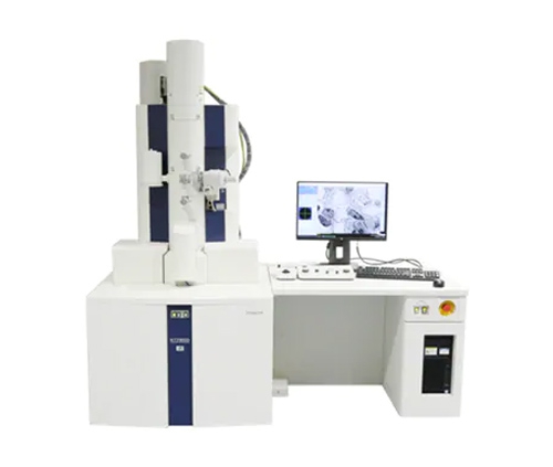
Large transmission electron microscope
Large-scale transmission electron microscopes (conventional TEM) generally use 80-300kV electron beam acceleration voltage. Different models correspond to different electron beam acceleration voltages. The resolution is related to the electron beam acceleration voltage and can reach 0.2-0.1nm. High-end models can achieve atomic Level resolution.
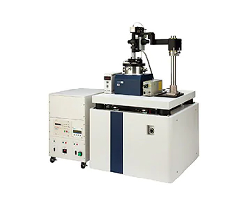
Low voltage transmission electron microscope
The electron beam acceleration voltage (5kV) used by the low-voltage electron microscope (LVEM) is much lower than that of the large-scale transmission electron microscope. A lower acceleration voltage will increase the intensity of the interaction between the electron beam and the sample, thereby improving the image contrast and contrast, which is especially suitable for samples such as polymer and biology. At the same time, the low-voltage transmission electron microscope will cause less damage to the sample.
Larger electron microscopes have low resolution, 1-2nm. Due to the low voltage, transmission electron microscope, scanning electron microscope and scanning transmission electron microscope can be integrated in one device
Cryo-electron microscopy
Cryo-microscopy is usually an ordinary transmission electron microscope with a sample freezing device to cool the sample to the temperature of liquid nitrogen (77K), which is used to observe temperature-sensitive samples such as proteins and biological sections. By freezing the sample, the damage of the electron beam to the sample can be reduced, the deformation of the sample can be reduced, and a more realistic sample morphology can be obtained.