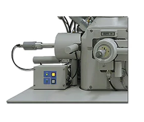
A microscope is an optical instrument composed of a lens or a combination of several lenses. It is also a symbol of people entering the atomic age. Microscope: Optical microscope and electron microscope: The optical microscope was created by Hooke and Levin Hooke. Microscopes can be divided into optical microscopes and electron microscopes. An optical microscope is usually composed of an optical part, an illumination part and a mechanical part. Obviously the optical part is important. The basic structural features of an electron microscope and an optical microscope are similar. But it is much stronger than the magnification and resolving power of the optical microscope. It uses electron flow as a new light source. Image the object. When using a microscope, the relationship between the magnification and other technical parameters must be coordinated according to the purpose of the microscopy and the actual situation. Get a good microscopic examination effect. So. How to use the transmission electron microscope correctly and safely. It is a subject. It is also a science. Let's discuss this issue below.
1. How to use transmission electron microscope
Before the experiment, place the microscope at a position slightly to the left of the table top in front of the seat. The lens holder should be within ten centimeters from the edge of the table: then turn on the light source switch and adjust the light intensity to the proper position: turn the nosepiece converter. Make the low-power lens face the light hole on the stage, first adjust the lens to about two centimeters from the stage, then look into the eyepiece with your left eye, and then adjust the height of the condenser. Adjust the aperture light to large. The light is incident into the lens barrel through the condenser. At this time, the field of view is in a bright state: place the glass slide to be observed on the stage, so that the observed part of the glass slide is located in the center of the light hole. Then clamp the glass slide with the specimen holder: first observe with a low power lens. Before observation. First turn the coarse focus adjustment handwheel. Raise the stage. The objective lens gradually approaches the glass slide. have to be aware of is. Do not allow the objective lens to touch the slide to prevent the lens from crushing the slide. Then. The left eye looks into the eyepiece. At the same time, do not close your right eye, and turn the coarse focus adjustment handwheel to slowly lower the stage. A magnified image of the material in the glass slide will soon be visible.
If the image seen in the field of view does not meet the experimental requirements. The moving handle of the stage can be adjusted slowly. When adjusting, it should be noted that the moving direction of the slide is exactly opposite to the moving direction of the object seen in the field of view. If the object image is not very clear. You can adjust the fine focus handwheel. Until the object image is clear: If you use a high magnification objective lens for further observation, it should be before switching the high magnification objective lens. Move the part of the object that needs to be magnified and observed to the center of the field of view. Generally a microscope with normal functions. The low magnification and high magnification objectives are basically parfocal. When the observation is clear with a low-power objective lens. The object image can still be seen by changing the objective lens of high magnification. But not necessarily very fresh. You can turn the fine focus adjustment handwheel to adjust: after switching the high magnification objective lens and seeing the object image clearly. The iris or condenser can be adjusted as needed. Make the light meet the requirements; after the observation, the objective lens should be removed from the light hole. Then restore the microscope. And check the parts for damage, and they can be put back to the original place after the inspection is completed.
2. Problems that should be paid attention to in the use of the microscope
The matching problem of the eyepiece and the objective lens: The eyepiece and the objective lens are the main optical parts of the microscope. Both can only be used correctly. In order to achieve good results. In order to match the eyepiece and objective lens correctly. The first thing to know is the logo on the objective lens housing. It is marked on the objective lens housing. The microscope is equipped with a set of eyepieces and a set of objective lenses. When using a microscope, in order to achieve high resolution, it is necessary to choose an eyepiece with a suitable magnification. when using it. The numerical aperture of the condenser should be equal to or close to the numerical aperture of the objective lens when the numerical aperture of the condenser is smaller than the numerical aperture of the objective lens used. The numerical aperture of the objective lens cannot be fully utilized. And reduce the image brightness and resolution.
2.1 Precautions for the use of optical microscope Microscope use: When using natural light source for microscopic examination, use a north-facing light source instead of direct sunlight: When using an artificial light source, use a fluorescent light source. When performing microscopy, the body should face the operating table and take an upright posture. The pupil distance of the eyepiece should be adjusted by the person, and the eyes should be opened naturally. Observe the specimen with the left eye. Observe the record and drawing with the right eye. At the same time adjust the focus with the left hand. Make the object image clear and move the specimen field of view. Right-handed recording and drawing. During microscopic examination, the specimen should be moved in a certain direction. Until the entire specimen is observed. In order not to miss the inspection and not to repeat. The heavy light of the microscope is the conversion of the light, the conversion of the objective lens and the adjustment of the light. When observing parasite specimens, light adjustment is very important. Objects in natural light. There are large and small, the color is dark and light, and some are colorless and transparent, and low-power and high-power objective lenses are converted more. Therefore, different specimens and requirements must be followed during microscopic examination. Need to adjust the focus and light at any time. Only in this way can the observed object image be clear.
2.2 On the light
Turn the low-power lens to the bottom of the lens barrel to be in line with the lens barrel: flip the mirror. Adjust to a bright field of view without shadows. The reflector has flat and concave sides. Use a flat surface when the light source is strong. Use concave when darker. When strong light is needed, raise the condenser and enlarge the aperture; when weak light is needed, lower the condenser or reduce the aperture appropriately: Place the specimen to be observed on the stage. Rotate the coarse adjuster to lower the lens barrel until the objective lens approaches the specimen and rotate the coarse adjuster at the same time. You must bend over the lens and carefully observe the distance between the objective lens and the specimen.

2.3 The use of the objective lens and the adjustment of the light
Microscopes generally have three objective lenses, namely low magnification, high magnification and oil lens. Fixed in the lens conversion disk L. When observing the specimen. First use a low-magnification objective lens. At this time, the field of view is larger and the specimen is easier to detect, but the magnification is smaller. Smaller objects are not easy to observe their structure. The magnification of high magnification objective lens is larger. Can observe tiny objects or structures using low and high magnification lenses. Such as when the object or its internal structure cannot be accurately identified under a low power lens. Then switch to high-power observation. Use oil glasses to observe. Generally, after adding a drop of oil, directly immerse the oil lens in the oil drop for microscopic observation.
2.4 How to use low magnification lens for high magnification lens
After the light is right. Move the thruster to look for the specimen that needs to be observed: If the volume of the specimen is large, its structure cannot be clearly seen and cannot be confirmed. Move the specimen to the center of the field of view. Then rotate the high magnification objective lens below the lens barrel: rotate the micro adjuster until the object image is clear: adjust the condenser and aperture to make the object image in the field of view clear.
2.5 matters needing attention
Before using the microscope. You should be familiar with the names of the various parts of the microscope and how to use them. In particular, the characteristics of the three types of objective lenses should be mastered: most of them are colorless and lighter in color. Therefore, attention must be paid to the adjustment of light: when fresh specimens are observed, a cover glass must be added to prevent the specimen from being dried and deformed due to evaporation or pollution and corroding the objective lens. At the same time, the specimen surface can be leveled and the light can be concentrated. Conducive to observation.
3. Maintenance of the microscope
When the microscope is taken out of the wooden box or packed. Hold the mirror arm tightly with your right hand. Hold the lens holder firmly with your left hand. Remove gently. Do not extract with only one hand to prevent the microscope from falling, and then gently place it on the practice table or put it in a wooden box: when the microscope is placed on the practice table. Put one end of the lens holder first. Then put all the mirror holders firmly. Do not allow the mirror holder to contact the table surface at the same time. This vibration is too large. The lens and micro adjuster are easily damaged: the microscope must be kept clean at all times. Do not allow oil and dust to adhere. If the lens is not clean, wipe it lightly with lens cleaning paper. If there is oil stains, first damp the lens cleaning paper with a little xylene to wipe it off: Do not pull out and remove the eyepieces and objective lenses casually. The mirror cover must be covered with a cloth to prevent dust from falling into the mirror cover. When changing the objective lens. After taking it down, place it upside down under a clean surface. And then put it into the wooden box placed in the tube of the objective lens.